Gonad Development
The aim of this project is to explore the reproductive biology of Bay of Fundy Striped Bass. Histological sectioning and the Gonadosomatic Index measure will be used.
Theoretically, the Gonadosomatic Index (GSI) provides a relative measure of gonad weight to fish body weight as an indicator of gonad maturity. The equation is:
GSI = (Gonad weights (g) /whole fish body weights (g)) *100
Histological sectioning provides direct evidence of individual cell growth including size and cell structure as a gonad develops.
Understanding reproductive biology is important to inform conservation actions, management and population health.
Objectives
- To characterize oocyte stages during gonad development.
- To quantify the proportion of oocyte stages at different gonad development stages.
- To estimate GSI seasonally in both sexes.
Methods
- Collect Striped Bass carcasses (not fillets) from recreational anglers, or scientifically sample Striped Bass.
- Record general information such as length and weight.
- Weigh gonad for GSI (if accurate fish weight is known).
- Extract a small piece of gonad and preserve in Bouin’s fixative, embed in paraffin wax, section, stain, and view under microscope.
Results
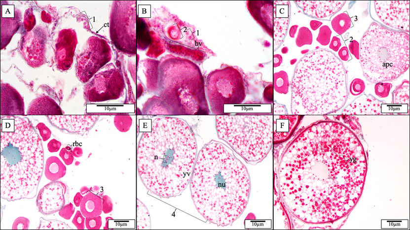
Female Gonads: Oocyte Development
So far, we have identified six stages of developing oocytes that eventually become eggs. The numbers corresponded to the stages of oocytes. Stage 1 oocyte, also called oogonia, can only be found under 40x magnification and has lots of associated connective tissue. Oogonia size dramatically increases at stage 2 cells and has a prominent nucleus located in the center (Panel B). Panel F is stage 6 cell for comparison.
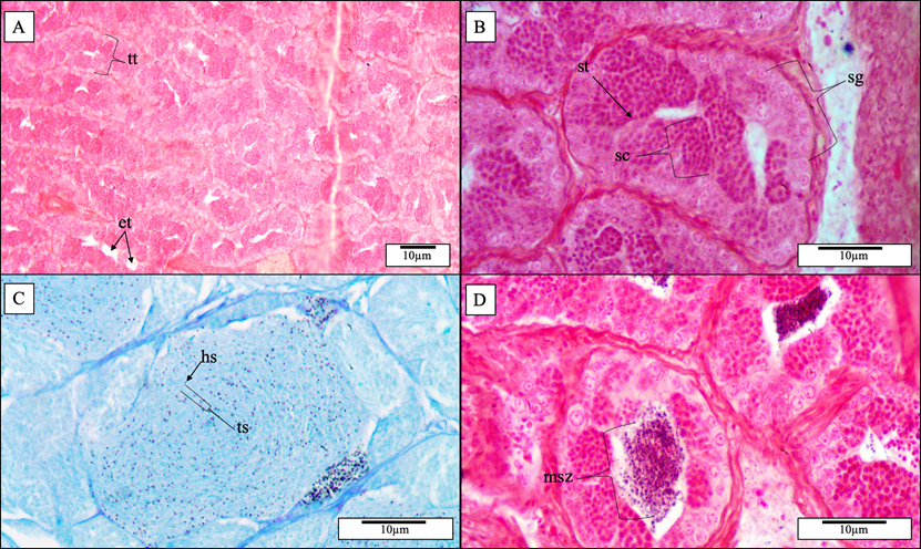
Male Gonads: Spermatogonium Development
Cell structure looks different in male and female gonads (bottom Panel). Male gonad tissue is more compact and has fewer developmental stages than females. Male takes four developmental stages for a spermatogonium to develop into sperm; this would be a mature gonad. Dark dots in Panel C and D corresponded to the head of a sperm. Outside of the spawning period, males were rarely seen to have mature sperm cells in testis tubules (white space in Panel A and B).
Project Supervision and Funding
This project is being undertaken by Zhe (Jackson) Yang under the co-supervision of Dr. Trevor Avery and Dr. Glenys Gibson. This project is funded by NSERC and Nova Scotia Transportation Infrastructure Renewal.